46 papers, an international user base, one open-source microscope: BrainSaw turns 10
5 October 2026
BrainSaw is a platform for high-throughput whole-brain microscopy, built a decade ago by the Sainsbury Wellcome Centre's Advanced Microscopy Facility. It has since been adopted by research groups worldwide, contributing to 46 published studies – from understanding the neurobiology of habits to unravelling the neural circuits that control escape.
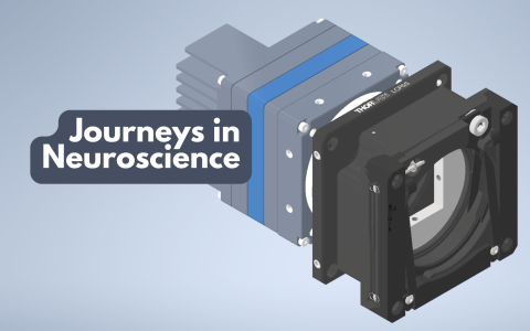
BrainSaw is an open-source serial two-photon tomography (STPT) system: it couples a multi-photon microscope to a vibratome and a three-axis stage. The tissue surface is exposed by the vibratome and scanned by the microscope, alternating between slicing and imaging until the full sample has been acquired. The data are then stitched together to create a 3D image.
STPT allows researchers to acquire high-resolution, whole-organ 3D image datasets – essential for neuroscience research but also used in cancer and immunology.
BrainSaw was developed by Dr Rob Campbell, Head of the Advanced Microscopy Facility at the Sainsbury Wellcome Centre (SWC). "Ten years ago, BrainSaw was something I built to solve a problem in our own lab – we wanted a system we could control, customise, and fix ourselves, something that was not possible with commercial alternatives. BrainSaw is by far the most heavily used platform in our Facility: researchers love the fact that they can acquire 3D whole brain datasets with minimal effort and no elaborate sample processing. It’s been amazing to watch the system not only be enthusiastically adopted in-house, but also now being used around the world," says Dr Campbell.
A new preprint on bioRxiv describes BakingTray, the acquisition software that drives SWC’s BrainSaw microscopes. This open-source software is highly modular, allowing adopters to build a BrainSaw STPT system that matches their needs.
"BakingTray has enabled us to rebuild an imaging system to make it robust and application-oriented, with tried-and-tested, user-friendly software that remains open to ad hoc modifications," said Dr Emmanuel Beaurepaire from the CNRS at École Polytechnique, France, a co-author of the preprint.
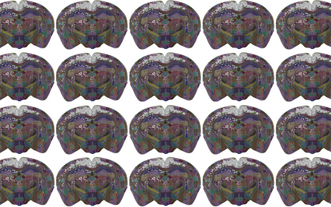
At SWC, the system is primarily used for electrode-track mapping, whole-brain cell counting, and mapping bulk axonal projections.
"Beyond the discoveries it enables, BrainSaw is a fantastic teaching example. We train early-career researchers to build two-photon microscopes themselves from scratch, giving them confidence in hands-on engineering skills that they'll take through their careers, whether they stay in this exact field or not. Skills like these enable our researchers to build their own tools if they can’t find an off-the-shelf solution," adds Dr Campbell.
Dr Alex Fratzl, former SWC PhD student, now a postdoc at IOB, Basel, said, "During my PhD, BrainSaw was a trustworthy daily companion. It made verifying injection sites and probe locations effortless, and watching the brain reveal its secrets in real time was one of the real joys of that period."
The SWC team hopes that more groups will use the platform, tailoring it for their own experiments and building a community of users.
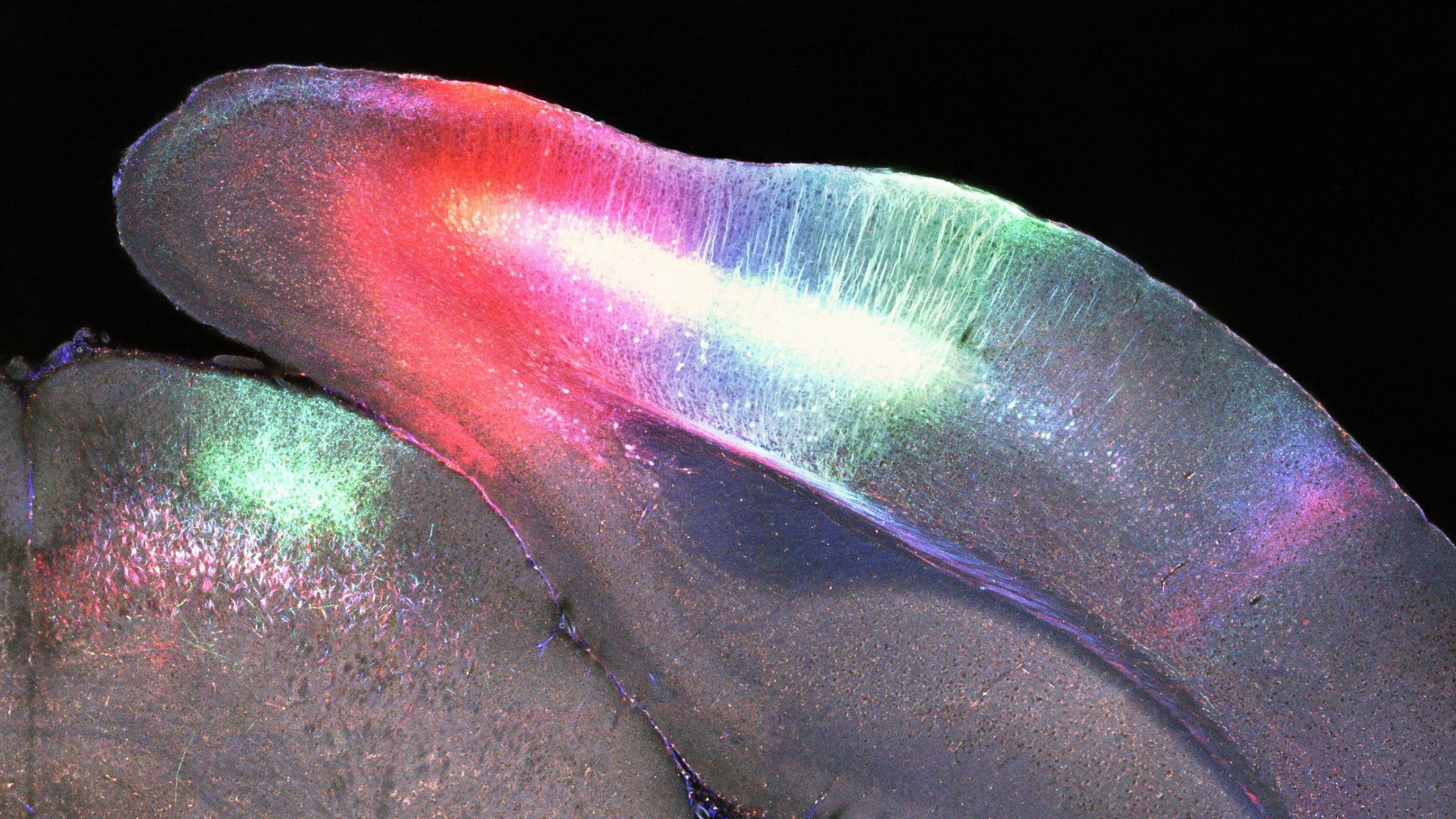
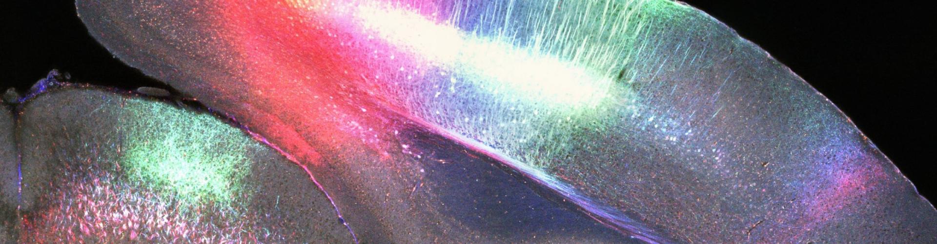
Mouse brain labelled with three AAV injections in layer 5 neurons of primary visual cortex: mCherry, GFP, and BFP imaged at 780 nm.
A proven platform, built to be replicated
BrainSaw’s workflow is automated and allows up to six samples to be imaged unattended, overnight, on a single microscope. Thousands of samples have been processed at SWC, and the system has proven to be highly robust. The team provides details that allow scientists to either build the system from scratch or adapt or upgrade existing systems or two-photon microscopes.
The system's components include:
· BrainSaw — CAD models and build instructions for assembling a serial-section two-photon microscope from scratch, or for adding serial-sectioning functionality to an existing system.
· BakingTray — modular MATLAB-based acquisition software, compatible with diverse hardware configurations, which acquires images via ScanImage.
· StitchIt — software that runs alongside BakingTray to pre-process data during acquisition, stream stitched sections in real time, and automatically assemble tile-scan data into complete sections once acquisition is finished.
· BrainGlobe — a suite of Python-based tools, maintained by SWC's Neuroinformatics team, for accessing, analysing and visualising the resulting anatomical data.
"BakingTray is on par with software of the top microscope companies, in terms of ease of use, stability, well-thought-out GUI and documentation. When one considers that it also controls a vibratome and is used for serial sectioning and imaging the whole brain, unattended, at cellular resolution, one starts to understand its power. On top of all that, having software that is robust, reliable and can image every labelled cell in the brain in about eight hours is what makes BT truly amazing. If you still want a cherry on the cake, it is open source," said Dr Rajeev Rajendran, a co-author of the preprint from the Netherlands Institute for Neuroscience.
Find out more:
Advanced Microscopy Facility Team:
Contact the SWC Advanced Microscopy Facility team
Media contact:
For more information or to speak to the researchers involved, please contact:
Alison Cranage, Research Communications and Engagement Manager, Sainsbury Wellcome Centre
E: a.cranage@ucl.ac.uk T: +44 (0)7917 922 068
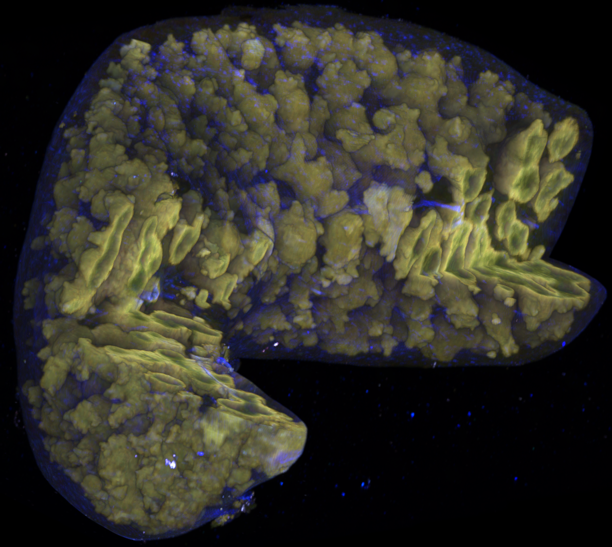
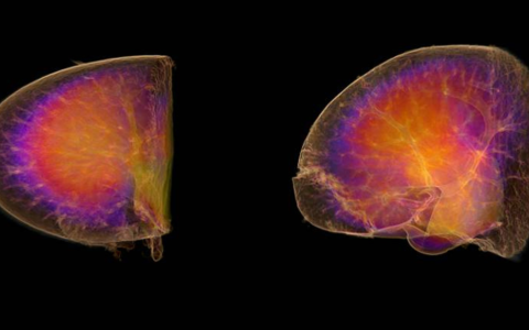
A 3D view of a mouse spleen with sparse confetti labelling.