
Muscle memories: how does sleep produce consolidation?
24 July 2026
By April Cashin-Garbutt
“The best bridge between despair and hope is a good night’s sleep” – E. Joseph Cossman.
Sleep not only makes us feel better, but it’s vital for helping us form memories and learn new skills. For many years, neuroscientists have studied how the brain consolidates episodic memories during sleep, which are memories of experiences such as what you did last weekend. But consolidation also occurs for procedural memories, so called “muscle memories”, such as learning to serve a tennis ball.
To understand how memories for motor skills are learned during sleep, Dr Emmett Thompson, a former Research Fellow in the Stephenson-Jones lab at SWC, designed a task where mice learned a sequence of steps by poking their nose into different ports. Just like a person learning a new dance, after a good night’s sleep the mice were much better at the task. Dr Thompson was eager to understand what was happening in the brain during sleep to produce this consolidation.
“We spend nearly half our lives sleeping, but little is known about what happens in the brain during sleep. Clearly sleep is fundamental to the way the brain works, and I am fascinated in studying its role in consolidating learning,” commented Dr Thompson.
The role of the hippocampus and the striatum in memory consolidation
Previous studies have shown that the hippocampus is very important for memory formation. Patient HM famously lost the ability to form new memories after his hippocampi were removed in an attempt to cure his severe epilepsy. For many years, the hippocampus has been thought of as the main organiser of all memory consolidation.
However, there have also been many parallel studies which have shown that motor skill learning is completely independent of the hippocampus. If you lesion the hippocampus, you can still learn motor skills normally, sometimes even faster.
“We wanted to test whether the hippocampus is really needed for all kinds of memory consolidation or whether there are separate parallel memory systems for episodic and procedural memories,” explained Dr Thompson.
The team looked at the striatum, a region of the brain known to be important for motor skill learning. By lesioning this area of the brain before and after learning, they observed that the dorsal striatum is essential for consolidating the nose poking memory task.
The lesioned mice still moved normally and tried the task, but they lost all memory of the correct sequence of movements. This implies that the striatum is involved in both forming and storing muscle memories.
Searching for replay during sleep
To look for reactivations of brain activity during sleep, known as replay, the team collaborated with PhD students at the Gatsby Computational Neuroscience Unit. Together they developed an unsupervised method that allowed them to decode the activity without making any prior assumptions.
“Studying brain activity during sleep is challenging as it is hard to tie activity patterns with movements as mice are not moving while they are asleep. The unsupervised method we developed looks for activity patterns when the mouse is awake and learns the repeating structures in these activities, called neural sequences.”
By mapping the neural sequences to behaviour during wake, the team were able to look for the same sequential patterns during sleep, thereby indirectly relating them to behaviour. This unsupervised method allowed them to observe replay of motor skill activity in the striatum.
“This is the first time anyone has found sequential neural replay of procedural activity in the striatum. We have found that this replay looks a lot like replay in other brain regions. The replay in the striatum is sequential, it recapitulates awake activity, and it also seems to be prioritised. This means that task-related activity is more likely to be replayed than non-task related activity, which was even true at a single cell level,” explained Dr Thompson.
To explore this prioritisation further, the researchers looked at how behaviour affects the content of replay and how the contents of replay affect changes in memory the next day. They showed that the brain gives priority to replaying patterns of activity linked to important experiences. Positive and negative outcomes affected these replay events in opposing ways.
When the team looked at the individual neurons, the ones that were more likely to be active during wake were also more likely to be boosted during sleep. Thus, the brain appears to be selecting for the neurons that are most involved in the task.
While the similarity of the replay in striatum and other brain regions suggests they might share a common mechanism, interestingly the team found that they do not. When they lesioned the hippocampus, the replay still occurred and there was no impact on learning. This suggests that replay is a common feature for memory consolidation, but the hippocampus is not.
“Our research shows that the current idea of the hippocampus as a central organiser that’s necessary for all types of memory consolidation cannot be true. Local brain regions are capable of consolidating independently and the hippocampus is not needed for learning memories with a single motor modality,” explained Dr Thompson.
Uncovering the source of hippocampus-independent replay
This research raises many questions including what triggers replay if not the hippocampus? Can neural circuits spontaneously produce this activity? And how is replay in different memory systems coordinated?
The Stephenson-Jones lab is looking to explore whether striatal replay and dopamine release is synchronised. “Previous work has shown that when the striatum is learning, dopamine is present. We want to look at our replay events while also recording dopamine activity in the striatum and see whether they correlate. This could help tell us what these replays mean,” concluded Dr Thompson.
Find out more
- Read the full paper in Nature Neuroscience, ‘Replay of procedural memory is independent of the hippocampus’ DOI: 10.1038/s41593-026-02362-5
- Learn more about research in the Stephenson-Jones Lab
Banner image
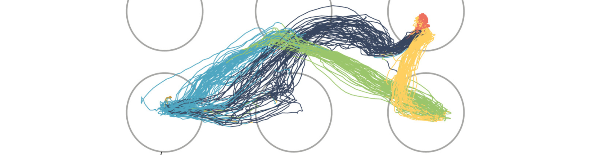
Banner image shows example movement tracking (animal head) in the five port task, coloured by the type of neural sequence. Credit: Stephenson-Jones Lab